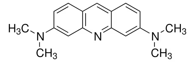
CAS Number494-38-2
SynonymsN,N,N',N'-Tetramethyl-3,6-acridinediamine; 3,6-Bis(dimethylamino)acridine Solvent Orange 15; Acridine Orange base; N,N,N',N'-Tetramethylacridine-3,6-diamine; N3,N3,N6,N6-Tetramethylacridine-3,6-diamine; euchrysine; ACRIDINE ORANGE
Molecular FormulaC17H19N3
Molecular Weight265.35
Purity75.00%
Density1.2±0.1 g/cm3
Boiling Point468.6±25.0 °C at 760 mmHg
Melting Point165 °C (dec.) (lit.)
Appearancepowder
| Chemical & Physical Properties | |
|---|---|
| CAS Number | 494-38-2 |
| Catalog No. | 2A-9009053 |
| Chinese Name | 吖啶橙 |
| Synonyms | N,N,N',N'-Tetramethyl-3,6-acridinediamine; 3,6-Bis(dimethylamino)acridine Solvent Orange 15; Acridine Orange base; N,N,N',N'-Tetramethylacridine-3,6-diamine; N3,N3,N6,N6-Tetramethylacridine-3,6-diamine; euchrysine; ACRIDINE ORANGE |
| Molecular Formula | C17H19N3 |
| Molecular Weight | 265.35 |
| Purity | 75.00 |
| Density | 1.2±0.1 g/cm3 |
| Boiling Point | 468.6±25.0 °C at 760 mmHg |
| Melting Point | 165 °C (dec.) (lit.) |
| Appearance | powder |
| Storage Condition | The warehouse is ventilated and dried at low tempe |
| Application | Acridine Orange is a cell-permeable fluorescent dye that stains organisms (bacteria, parasites, viruses, etc.) bright orange and, when used under appropriate conditions (pH=3.5, Ex=460 nm), distinguis |
| PubChem ID | 24854106 |
| MDL Number | MFCD00005029 |
| SMILES | CN(C)c1ccc2cc3ccc(cc3nc2c1)N(C)C |
| InChI | 1S/C17H19N3/c1-19(2)14-7-5-12-9-13-6-8-15(20(3)4)11-17(13)18-16(12)10-14/h5-11H,1-4H3 |
| InChI Key | DPKHZNPWBDQZCN-UHFFFAOYSA-N |
| NACRES Code | NA.47 |
| UNSPSC | 12171500 |
| MSDS | msds/9053_1_494_38_2.pdf |
| Bioactivity | Acridine Orange base is a cell-permeable fluorescent dye that stains organisms (bacteria, parasites, viruses, etc.) bright orange and, when used under appropriate conditions (pH=3.5, Ex=460 nm), distinguishes human cells in green for detection by fluorescence microscopy. Acridine Orange base fluoresces green when bound to dsDNA (Ex=488, Em=520-524) and red when bound to ssDNA (Ex=457, Em=630-644) or ssRNA (Ex=457, Em=630-644), also can be used in cell cycle assays[1][2][3]. Related Catalog Research Areas >> Others Signaling Pathways >> Others >> Others In Vitro Guidelines (Following is our recommended protocol. This protocol only provides a guideline, and should be modified according to your specific needs). 1. Differential staining of DNA and RNA of unfixed cells[1]: (1) Set up the flow cytometer with excitation at 488 nm, using emission filters and a dichroic mirror that discriminate green fluorescence (measured at 515-545 nm) and red luminescence (measured preferably above 640 or 650 nm). (2) Transfer a 0.2-mL aliquot of the original cell suspension to a small glass or plastic tube (e.g., 2- or 5-mL volume). Chill on ice. (3) Gently add 0.4 mL ice-cold cell permeabilizing solution. Wait 15 s, keeping cells on ice. (4) Gently add 1.2 mL ice-cold Acridine Orange base staining solution. Keep cells on ice in the dark. (5) Measure and record cell fluorescence in the flow cytometer during the 2 to 10 min after addition of Acridine Orange base staining solution. 2. Differential staining of fixed cells[1]: (1a) For cells in suspension culture or hematologic samples: Rinse cells once with ice-cold PBS and suspend in ice-cold PBS at ∼106 cells/mL. (1b) For cells attached to tissue culture plates: Collect cells from flasks or petri plates by trypsinization, pool the trypsinized cells with cells floating in the medium (mostly detached mitotic and dead cells), and rinse once with medium containing serum to inactivate the trypsin. Suspend cells in ice-cold PBS at ∼106 cells/mL. (1c) For cells isolated from solid tumors: Rinse cells free of any enzyme used for cell dissociation and suspend in ice-cold PBS at ∼106 cells/mL. (2) With a Pasteur pipet transfer 1 mL cell suspension to a 15-mL conical glass tube containing 10 mL ice-cold 70% ethanol. Fix cells ≥2 h on ice. (3) Centrifuge tubes 5 min at 300 × g, 4℃. Remove all ethanol, rinse cells once with ice-cold PBS, and suspend in ice-cold PBS at a density of < 2 × 10⁶ cells/mL. (4) Withdraw 0.2 mL cell suspension (≤ 2 × 10⁵ cells) and transfer to a small tube (e.g., 2 or 5 mL volume). Chill on ice. (5) Add 0.4 mL ice-cold permeabilizing solution. Wait 15 s, keeping cells on ice. (6) Add 1.2 mL ice-cold Acridine Orange base staining solution. Keep cells on ice. (7) Measure and record cell fluorescence in the flow cytometer during the 2 to 10 min after addition of Acridine Orange base staining solution. References [1]. Darzynkiewicz Z, et al. Differential staining of DNA and RNA. Curr Protoc Cytom. 2004 Nov;Chapter 7:Unit 7.3. [2]. Mirrett S. Acridine orange stain. Infect Control. 1982 May-Jun;3(3):250-2. [3]. Yektaeian N, et al. Lipophilic tracer Dil and fluorescence labeling of acridine orange used for Leishmania major tracing in the fibroblast cells. Heliyon. 2019 Dec 18;5(12):e03073. Chemical & Physical Properties Density 1.2±0.1 g/cm3 Boiling Point 468.6±25.0 °C at 760 mmHg Melting Point 165ºC (dec.)(lit.) Molecular Formula C17H19N3 Molecular Weight 265.353 Flash Point 237.2±23.2 °C Exact Mass 265.157898 PSA 19.37000 LogP 4.65 Vapour Pressure 0.0±1.2 mmHg at 25°C Index of Refraction 1.710 InChIKey DPKHZNPWBDQZCN-UHFFFAOYSA-N SMILES CN(C)c1ccc2cc3ccc(N(C)C)cc3nc2c1 |
| Use of | Acridine Orange base is a cell-permeable fluorescent dye that stains organisms (bacteria, parasites, viruses, etc.) bright orange and, when used under appropriate conditions (pH=3.5, Ex=460 nm), distinguishes human cells in green for detection by fluorescence microscopy. Acridine Orange base fluoresces green when bound to dsDNA (Ex=488, Em=520-524) and red when bound to ssDNA (Ex=457, Em=630-644) or ssRNA (Ex=457, Em=630-644), also can be used in cell cycle assays[1][2][3]. Properties Name acridine orange Synonym More Synonyms Biological Activity Description Acridine Orange base is a cell-permeable fluorescent dye that stains organisms (bacteria, parasites, viruses, etc.) bright orange and, when used under appropriate conditions (pH=3.5, Ex=460 nm), distinguishes human cells in green for detection by fluorescence microscopy. Acridine Orange base fluoresces green when bound to dsDNA (Ex=488, Em=520-524) and red when bound to ssDNA (Ex=457, Em=630-644) or ssRNA (Ex=457, Em=630-644), also can be used in cell cycle assays[1][2][3]. Related Catalog Research Areas >> Others Signaling Pathways >> Others >> Others References [1]. Darzynkiewicz Z, et al. Differential staining of DNA and RNA. Curr Protoc Cytom. 2004 Nov;Chapter 7:Unit 7.3. [2]. Mirrett S. Acridine orange stain. Infect Control. 1982 May-Jun;3(3):250-2. [3]. Yektaeian N, et al. Lipophilic tracer Dil and fluorescence labeling of acridine orange used for Leishmania major tracing in the fibroblast cells. Heliyon. 2019 Dec 18;5(12):e03073. Chemical & Physical Properties Molecular Formula C17H19N3 Exact Mass 265.157898 PSA 19.37000 Index of Refraction 1.710 InChIKey DPKHZNPWBDQZCN-UHFFFAOYSA-N |
| Transport Info | Product Name: Acridine Orange |
| Related Products in Specialty Chemicals | ||
|---|---|---|
| Achromycin hydrochloride | 64-75-5 | 2A-9009045 |
| Acipimox | 51037-30-0 | 2A-9009047 |
| Acitretin | 55079-83-9 | 2A-9009048 |
| Acoxatrine | 748-44-7 | 2A-9009051 |
| Acridine | 260-94-6 | 2A-9009052 |
| Acrinathrin | 101007-06-1 | 2A-9009054 |
| Ethacridine lactate monohydrate | 6402-23-9 | 2A-9009055 |
| Acrivastine | 87848-99-5 | 2A-0202277 |
| Actarit | 18699-02-0 | 2A-9009057 |
| Adibendan | 100510-33-6 | 2A-9009062 |
| Browse all Specialty Chemicals products » | ||
Phone:
630-322-8886
630-322-8887
Email:
Office Locations:
Chicago, IL, USA
San Francisco, CA, USA


 EN
EN


 SDS
SDS